Large Animal Dissection - The Neck
VMED 5125 - Basic and Applied Veterinary Anatomy - III Spring Semester. Large Animal Dissection - The Neck - Part I. D. J. Hillmann, D. V. M. , Ph. D. Professor, Veterinary Gross Anatomy. The Dog (Review). The jugular groove. Note how deep. The Ox (bovine). The Horse. Cutaneous mm. & subcutaneous bursae of the Head and Trunk of the Horse. Muscles of the Neck of the Dog (Review). M. brachiocephalicus M. omotransversarius M. sternocephalicus. Muscles of the Neck of the Ox. (Viewed from the Lateral Aspect). Muscles of the Neck of the Horse. M. pectoralis superficialis M. pectoralis profundus M. subclavius (absent). Pectoral Musculature of the Dog (Review). M. pectoralis superficialis M. pectoralis profundus M. subclavius. Pectoral Musculature of the Ox. Pectoral Musculature of the Horse. (Viewed from the Cranial Aspect or Looking Caudally).
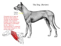
The jugular groove clearly lies between the cleidocephalicus (dorsally) and sternocephalicus (ventrally) at the caudal end. Cranially it is less distinct, as the sternocephalicus crosses deep to the vein. There is no sternomandibularis to continue the ventral border of the groove. (See muscles of the neck!).
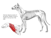
Note how deep the location of the cervical vertebral column is relative to the top-line of the neck.
The jugular groove between the cleidocephalicus (dorsolaterally) and the sternomandibularis (ventro-medially) is rather wide in the ox. The sternomastoideus forms the deep wall of the groove and separates the external jugular vein from the carotid artery except for a small area near the first rib, where there is no muscle between the two vessels. (See muscles of the neck!).
- Pet Presentations
- MS PowerPoint 7868 KB
- 2020 m.
- English
- 14 pages (456 words)
- University
- Roberta


